
Imagine the blood flow in your body as a highway system. Now imagine that one of the main arteries leading into your liver gets blocked by a clot. That is exactly what happens with Portal Vein Thrombosis (PVT), a condition where a blood clot forms in the portal vein, the large vessel carrying nutrient-rich blood from the intestines to the liver. It sounds scary, and it is serious, but here is the good news: if you catch it early and treat it correctly, the outcomes are often excellent. In fact, patients who start treatment quickly have an 85% five-year survival rate.
The key to surviving and thriving with PVT lies in two words: diagnosis and anticoagulation. You need to know how doctors spot this blockage before it causes permanent damage, and you need to understand which blood-thinning medications work best for your specific situation. Whether you have cirrhosis or not, the rules change slightly, so let’s break down what you actually need to know.
How Doctors Spot Portal Vein Thrombosis
You might not feel anything at first. Many people with PVT have no symptoms until the clot grows large enough to cause pressure issues. When symptoms do appear, they often include abdominal pain, swelling in the belly (ascites), or confusion due to toxins building up because the liver isn't filtering blood properly. If the clot blocks blood flow to the intestines, it can cause severe pain and vomiting-a medical emergency known as mesenteric ischemia.
So, how do we find it? The first line of defense is usually an ultrasound. Specifically, a Doppler ultrasound. This test is non-invasive and highly effective, detecting portal vein abnormalities with about 89-94% sensitivity. The sonographer looks for three things:
- Absence of blood flow: Normally, blood should move smoothly toward the liver. If it stops or reverses, that’s a red flag.
- Echogenic material: The clot itself shows up as a bright spot inside the dark vein on the screen.
- Cavernous transformation: In chronic cases, the body tries to bypass the blockage by creating new, tiny collateral veins. These look like a network of small vessels around the original vein.
If the ultrasound is unclear, doctors will move to a CT scan or MRI with contrast. These provide a detailed map of the clot’s size and location. They classify the blockage as minimally occlusive (less than 50% blocked), partially occlusive (50-99%), or completely occlusive (100%). Knowing the severity helps decide how aggressive the treatment needs to be.
The Role of Anticoagulation in Treatment
Once the clot is confirmed, the goal is clear: stop it from growing, dissolve it if possible, and prevent new clots. This is where anticoagulation comes in. Think of anticoagulants not as "clot busters" (which actively dissolve clots) but as agents that prevent the clot from getting bigger while your body’s natural systems slowly break it down. This process is called recanalization.
Timing is everything. Studies show that if you start anticoagulation within six months of diagnosis, you have a 65-75% chance of complete resolution. Wait longer, and that rate drops to just 16-35%. For acute cases, starting treatment immediately is standard care, even if you have cirrhosis, provided your bleeding risk is managed.
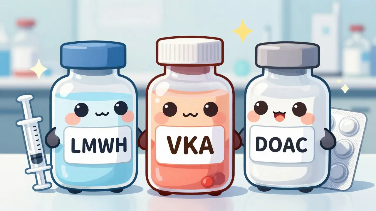
There are three main types of anticoagulants used today, each with pros and cons:
- Low Molecular Weight Heparin (LMWH): Drugs like enoxaparin are injected under the skin. They are often the first choice for initial treatment because their effect is predictable and they don’t require frequent blood monitoring. Typical dosing is weight-based, such as 1 mg/kg twice daily.
- Vitamin K Antagonists (VKAs): Warfarin is the classic oral pill. It requires regular blood tests (INR checks) to ensure the dose is right. The target INR is usually between 2.0 and 3.0. It’s effective but tricky to manage because diet and other drugs affect its potency.
- Direct Oral Anticoagulants (DOACs): Newer pills like rivaroxaban and apixaban are easier to use. No routine blood monitoring is needed. Recent data suggests they may offer higher recanalization rates than warfarin in non-cirrhotic patients.
| Drug Class | Examples | Recanalization Rate (Non-Cirrhotic) | Monitoring Required | Bleeding Risk Profile |
|---|---|---|---|---|
| LMWH | Enoxaparin | 55-65% | Anti-Xa levels (optional) | Moderate; reversible with protamine |
| VKA | Warfarin | 40-50% | Weekly INR checks initially | Variable; depends on INR control |
| DOACs | Rivaroxaban, Apixaban | 65-75% | None routinely | Low-Moderate; reversal agent available |
Treatment Differences: Cirrhosis vs. Non-Cirrhosis
Your liver health changes the game entirely. If you do not have cirrhosis, the focus is purely on dissolving the clot and finding out why it formed. About 25-30% of non-cirrhotic PVT cases are linked to thrombophilias-genetic conditions that make your blood prone to clotting. In these cases, you might need lifelong anticoagulation.
If you have cirrhosis, the situation is more delicate. Your liver is already scarred, and your blood vessels are fragile. The biggest fear is variceal bleeding-bleeding from enlarged veins in the esophagus or stomach. Before starting any blood thinner, doctors must screen for these varices using an endoscopy. If high-risk varices are found, they will band them first. This simple step reduces major bleeding events significantly, from 15% down to 4% in some studies.
For cirrhotic patients with Child-Pugh A or B disease (mild to moderate liver dysfunction), anticoagulation is safe and recommended. However, for those with Child-Pugh C disease (severe dysfunction) or uncontrolled ascites, the bleeding risk often outweighs the benefits, and anticoagulation may be contraindicated.
When Blood Thinners Aren't Enough
Sometimes, despite medication, the clot doesn't go away, or it causes severe complications like intestinal ischemia. In these cases, doctors consider interventional procedures.
TIPS (Transjugular Intrahepatic Portosystemic Shunt): This procedure creates a channel inside the liver to bypass the blockage, lowering pressure in the portal vein. It has a 70-80% technical success rate but carries a risk of hepatic encephalopathy (brain fog due to toxins) in 15-25% of patients. It’s usually reserved for cases where anticoagulation fails or portal hypertension is severe.
Thrombectomy: In emergencies, doctors may use catheters to mechanically remove the clot. This offers immediate relief but is only available at specialized centers and has a higher complication rate.

Long-Term Management and Follow-Up
Treating PVT is a marathon, not a sprint. How long do you stay on blood thinners?
- Provoked PVT: If the clot was caused by a temporary factor (like surgery or infection), you typically take anticoagulants for at least 6 months after the trigger is gone.
- Unprovoked or Genetic PVT: If you have a genetic clotting disorder or active cancer, you may need lifelong anticoagulation.
- Cirrhotic PVT: Often treated indefinitely unless the patient undergoes a liver transplant.
Regular follow-up ultrasounds are crucial. They check if the vein is opening up (recanalizing) and monitor for new clots. Don’t skip these appointments. Early detection of recurrence means easier treatment.
Living With Portal Vein Thrombosis
Life with PVT requires vigilance but doesn’t have to limit you. Here are practical tips:
- Stay Hydrated: Dehydration thickens the blood, increasing clot risk. Drink plenty of water.
- Avoid NSAIDs: Painkillers like ibuprofen or naproxen can increase bleeding risk and harm the liver. Use acetaminophen (paracetamol) cautiously, following your doctor’s dosage limits.
- Dietary Adjustments: If you have cirrhosis, limit salt to reduce fluid buildup. Avoid alcohol completely.
- Know the Warning Signs: Seek immediate help if you experience black tarry stools, vomiting blood, sudden severe abdominal pain, or confusion.
The landscape of PVT treatment is improving rapidly. New guidelines from organizations like the American Association for the Study of Liver Diseases (AASLD) now support the use of DOACs in more patients, including some with compensated cirrhosis. Reversal agents like andexanet alfa are also available now, making DOACs safer in case of emergency bleeding.
Remember, PVT is manageable. With early diagnosis, the right anticoagulant, and careful monitoring, most people recover well. Work closely with your hepatologist, ask questions, and don’t hesitate to seek a second opinion if you feel uncertain about your treatment plan.
Is Portal Vein Thrombosis fatal?
If left untreated, PVT can lead to serious complications like intestinal death or severe liver failure, which can be fatal. However, with early diagnosis and proper anticoagulation therapy, the 5-year survival rate is approximately 85%. The key is timely intervention.
Can Portal Vein Thrombosis resolve on its own?
It is rare for a significant portal vein thrombus to resolve completely without treatment. While the body can sometimes form collateral vessels to bypass the blockage (cavernous transformation), this does not fix the underlying clot and often leads to chronic portal hypertension. Anticoagulation is necessary to promote true recanalization.
Are DOACs safe for patients with cirrhosis?
DOACs are generally considered safe for patients with compensated cirrhosis (Child-Pugh A or selected B7 patients). However, they are contraindicated in decompensated cirrhosis (Child-Pugh C) due to increased bleeding risks and altered drug metabolism. Always consult a hepatologist before starting DOACs if you have liver disease.
How long do I need to take blood thinners for PVT?
The duration depends on the cause. For provoked PVT (e.g., post-surgery), treatment is typically 6 months. For unprovoked PVT or those with genetic clotting disorders, lifelong anticoagulation is often recommended. Patients with cirrhosis may require indefinite treatment unless they receive a liver transplant.
What are the side effects of anticoagulation for PVT?
The primary risk of anticoagulation is bleeding. Minor side effects include bruising easily and nosebleeds. Major bleeding events occur in 5-12% of cirrhotic patients and 2-5% of non-cirrhotic patients. Regular monitoring and screening for varices help mitigate these risks.
Gareth Tyler
May 31, 2026 AT 11:18good read man really helps to understand the basics without getting lost in medical jargon
Sharon O’Mahonh
June 2, 2026 AT 08:59i think it is crucial for us to recognize that anticoagulation therapy isn't just about stopping a clot but actually facilitating recanalization which is a complex physiological process where the body naturally breaks down the thrombus over time while preventing further propagation so we must view these medications as facilitators of natural healing rather than mere suppressants of pathology and this shift in perspective can greatly empower patients to adhere to their treatment regimens with greater confidence and understanding of the underlying mechanisms at play in their vascular system
Jonhnnie john13
June 2, 2026 AT 13:09the data presented here is superficial at best ignoring the significant heterogeneity in patient responses to DOACs versus LMWH especially in those with underlying cirrhosis where pharmacokinetic alterations are profound and often unpredictable making the blanket recommendation of DOACs potentially dangerous without rigorous individualized assessment of hepatic reserve and bleeding risk profiles which are rarely captured in such broad strokes
Anthony Padilla
June 3, 2026 AT 02:52hey everyone i wanted to share that in my country we often see different protocols for PVT management depending on access to specialized care so its important to remember that what works in one healthcare system might not be feasible everywhere else and we should always encourage open dialogue between patients and doctors to find the best path forward for each unique situation because health is deeply personal and cultural context matters a lot when discussing long term treatments like this
Elizabeth Fandry
June 3, 2026 AT 05:45One must consider the existential weight of such a diagnosis 🧠 The portal vein is not merely a vessel but a conduit of life itself, and its obstruction speaks to the fragility of our biological systems. To reduce this to simple statistics is to ignore the profound philosophical implications of living with a compromised circulatory architecture. We are but vessels ourselves, subject to the whims of coagulation and flow. 🩸
Madeline Petes
June 3, 2026 AT 21:19i was so scared when i got diagnosed but starting on apixaban changed everything for me and now i feel like myself again so if you are reading this dont lose hope because there are real options out there that work and you deserve to live your life fully without letting fear dictate your choices every single day
Ramanath Rao
June 5, 2026 AT 10:05This article is completely misleading! You cannot generalize treatment protocols across all demographics without considering genetic predispositions common in South Asian populations where Factor V Leiden is less prevalent but other thrombophilias are more common. Your failure to address these specific ethnic variations renders your advice useless and potentially harmful to millions who rely on accurate medical information. Wake up and do better research!
Russell Russell
June 5, 2026 AT 12:34it is inspiring to see how far medicine has come in managing conditions like PVT and i believe that with the right mindset and support system anyone can overcome these challenges so let us continue to share knowledge and uplift each other on this journey towards better health and wellness because together we are stronger and more informed
Cyburg Adeoye
June 6, 2026 AT 11:29I would like to emphasize the importance of multidisciplinary care teams; gastroenterologists, hematologists, and hepatologists must collaborate seamlessly to optimize outcomes for patients with portal vein thrombosis. Furthermore, the integration of advanced imaging modalities such as contrast-enhanced ultrasound and MRI elastography can provide nuanced insights into liver stiffness and collateral formation, thereby guiding more precise therapeutic interventions and improving overall patient stratification and prognostic accuracy.
Joseph Teichman
June 6, 2026 AT 12:19stick to the plan and stay hydrated guys